
Field Notes · October 8, 2025 · 6 min · By Fletcher Imafidon
What is Mohs surgery, and why does it have the highest cure rate?
The technique that checks the margins under the microscope while you wait.
Mohs micrographic surgery is the gold standard for removing many skin cancers, and its advantage comes down to one thing: it checks the edges of the tumor under the microscope before you leave.
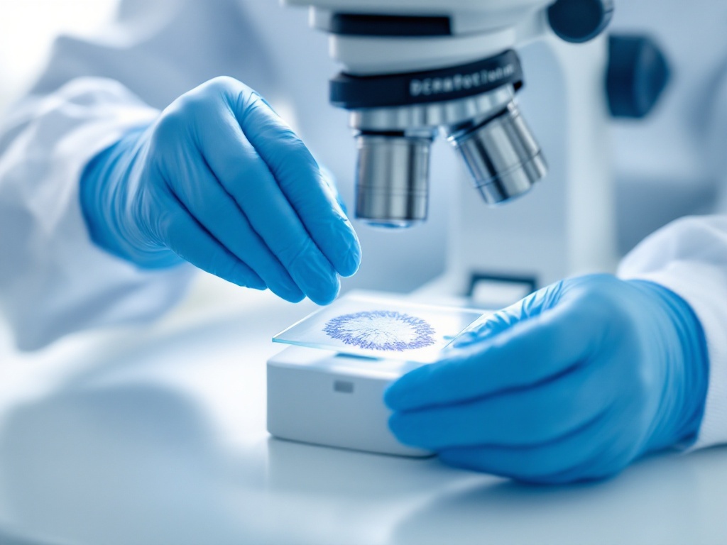
In a standard excision, the surgeon removes the visible cancer plus a margin and sends it to a lab; results come days later, and if the margins are not clear, you return for more surgery. In Mohs, the surgeon removes a thin layer, maps it precisely, and examines essentially the entire margin under a microscope on-site. If cancer remains at an edge, the surgeon removes only more tissue from that exact spot and rechecks, repeating until the margins are clear, all in one visit.
Two benefits follow. The cure rate is the highest of any treatment for common skin cancers, often around 99 percent for primary basal and squamous cell carcinomas, because no tumor is left behind unseen. And because tissue is removed only where cancer actually is, Mohs spares the most healthy skin, which matters enormously on the face. That combination of precision and tissue conservation is why Mohs is the standard for skin cancer in cosmetically and functionally sensitive areas.
Related reading: What to expect on the day of Mohs surgery.
Further reading: Who should have Mohs micrographic surgery? (Curr Opin Otolaryngol Head Neck Surg 2010); Mohs Micrographic Surgery: A Brief Overview (Semin Plast Surg 2024); Frederic Mohs: A Trailblazer in Dermatologic Surgery (Cureus 2024).